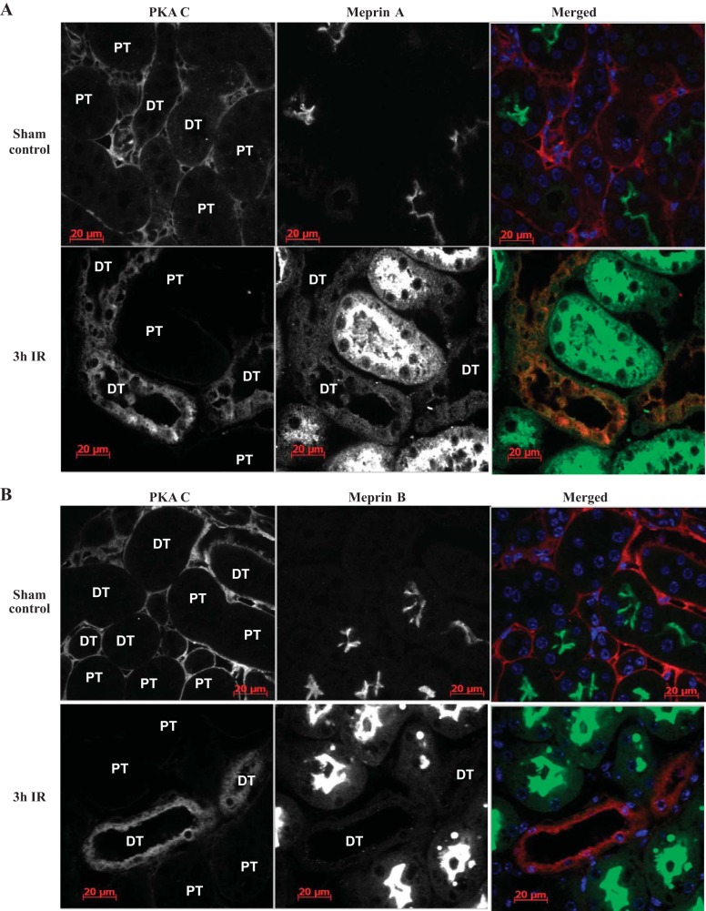
Fig. 9.
Immunolocalization of meprins (green) and PKA C (red) in the tubules of kidney tissue from WT mice with ischemia-reperfusion (IR)-induced renal injury. IR renal injury was induced in 12-wk-old male mice by surgical clamping of renal arteries for 26 min. The mice were euthanized at 3 h post-IR, and the tissue was paraffin embedded. Immunofluorescence staining with anti-meprin antibodies (green) and anti-PKA C antibodies (red) was used to localize the proteins, and imaged by confocal microscopy. DAPI was used to stain the nuclei (blue). Expression of meprins was restricted to the brush-border membrane of proximal tubules (PT) in sham-operated control mice, and the PKA C expression pattern was comparable in proximal and distal tubules (DT) in kidney tissue. However, at 3 h post-IR, meprins were redistributed to the cytosol of proximal tubule cells, and the levels of PKA C were lower in the meprin-expressing proximal tubules compared with the distal tubules (lacking meprins).

