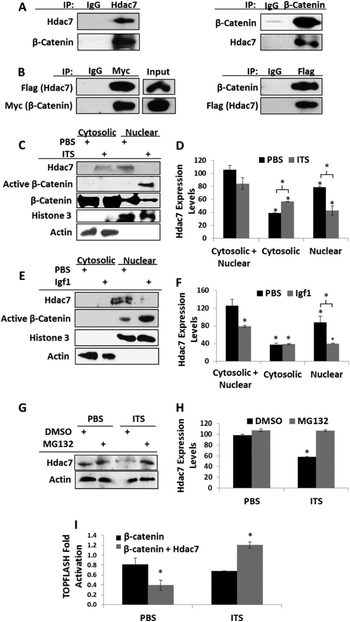
FIGURE 4.

Chondrogenic stimuli promote Hdac7 degradation and β-catenin activity. A, co-immunoprecipitations of Hdac7 and β-catenin endogenous to ATDC5 cells. B, ATDC5 cells were transfected with Flag-tagged Hdac7 and Myc-tagged β-catenin. Co-immunoprecipitations were performed as shown. C and D, ATDC5 cells were serum starved, treated with 1× ITS for 24 h, and then biochemically fractionated into cytosolic and nuclear extracts. C, Western blotting for the indicated proteins was performed and D, the expression levels of Hdac7 in each compartment were quantified, n = 3. E and F, ATDC5 cells were serum starved, treated with 10 ng/ml Igf1 for 24 h, and then biochemically fractionated into cytosolic and nuclear extracts. E, Western blotting for the indicated proteins was performed and F, the expression levels of Hdac7 in each compartment were quantified, n = 3. G and H, ATDC5 cells were serum starved in the presence of 5 nm MG132 or vehicle control. Cells were then treated with 1× ITS for 24 h. G, Western blotting was then performed as indicated and H, the expression levels of Hdac7 were quantified, n = 3. I, ATDC5 cells were transfected with the TOPFLASH reporter, a constitutively active form of β-catenin and pcDNA3 or pcDNA3-Hdac7 expression constructs as indicated. After transfection, cells were exposed to 1× ITS for 48 h. *, p < 0.05 compared with control, n = 3.
