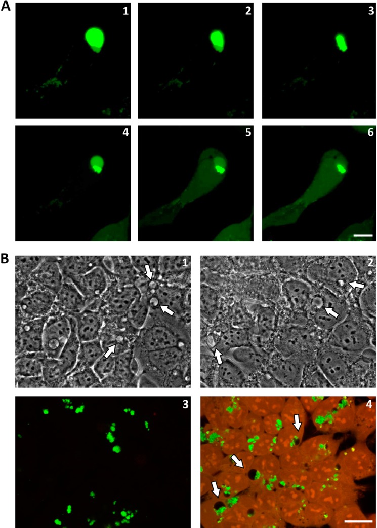
FIGURE 3.
Morphological analysis of enlarged vesicles. A, an HEK-293 cell bearing an enlarged vesicle containing a PepL aggregate was illuminated constantly with the confocal laser (argon, 488 nm) for 15 min. Morphological changes in the vesicle were followed by time lapse confocal microscopy: 30 s (1), 3 min (2), 9 min (3), 13 min (4), 14 min (5), and 15 min (6). B, fixation artifacts. HEK-293 cells were incubated for 24 h with PepL-DyLight 488 aggregates and imaged by bright field microscopy in vivo (1), bright field after fixation in 4% formaldehyde for 20 min (2), and confocal microscopy after fixation in 4% formaldehyde (3) or 2.5% glutaraldehyde (4), followed by permeabilization in 0.1% Triton X-100. Green, PepL; red, autofluorescence. Enlarged vesicles are indicated by arrows. Scale bar, 10 μm.

