FIGURE 2.
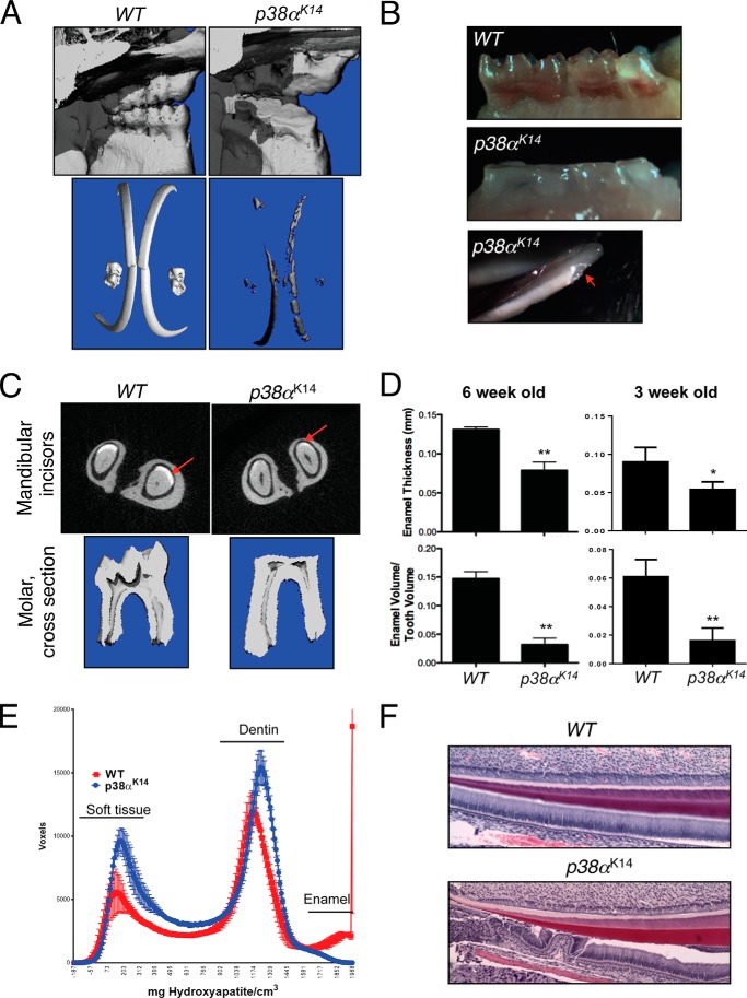
Dental phenotype of p38αK14 mice. A, skulls of 6-week-old p38αK14 and WT mice were scanned by μCT. Displayed are the lingual surfaces of the molars (top panels) and three-dimensional reconstructions windowed so that only enamel densities are visible (bottom panels). B, pictures of the molars from 3-week-old p38αK14 (middle panel) and WT mice (top panel) showing complete absence of molar cusps in p38αK14 mice. A view of the mandibular incisors shows spontaneous fracture of the enamel in p38αK14 mice (red arrow, bottom panel). C, the relative enamel volume and thickness of the indicated 6-week-old mice was determined by μCT analysis (top panels). Views of the two-dimensional slices from μCT analysis of p38αK14 and control mandibular incisors, showing a decrease in enamel thickness (bottom panels). D, quantitation of enamel thickness (mm) and enamel volume relative to total tooth volume in the mandibular incisors of 6- or 3-week-old p38αK14 and WT mice. ** indicates p < 0.01 by a two-tailed unpaired Student's t test. *, indicates p < 0.05. E, histogram plotting density on the x-axis versus the volume in voxels corresponding to density in mandibular incisors from 6-week-old p38αK14 and WT mice. The three peaks present correspond to soft tissue, dentin, and enamel densities from left to right. F, hematoxylin and eosin staining of 3-day-old mandibular incisors showing dissociation of soft tissue from dentin.

