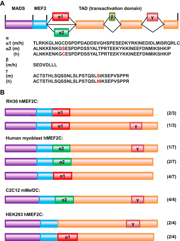
FIGURE 1.
MEF2C isoforms in muscle and RMS. A, schematic of the MEF2C isoforms. The sequences of the exons are indicated below. m, murine sequence; h, human sequence. Amino acids that differ among the species are shown in red. B, MEF2C isoforms identified in the indicated cell lines. The number beside each isoform indicates the number of individual isoform clones identified/total number of clones recovered.

