FIGURE 1.
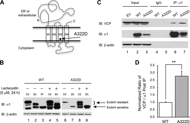
VCP interacts stronger with α1(A322D) subunits than with WT α1 subunits in HEK293 cells stably expressing WT α1β2γ2 or α1(A322D)β2γ2 GABAA receptors. A, topology of the α1 subunit. The A322D mutation is in the TM3 domain, labeled by an asterisk. B, effect of lactacystin on α1 subunit variant maturation. Treatment with lactacystin (5 μm, 24 h), a potent proteasome inhibitor, does not increase the endo H-resistant post-ER glycoform of the misfolding-prone α1(A322D) subunit in HEK293 cells (n = 2). Endo H-resistant α1 subunit bands represent properly folded, post-ER α1 subunit glycoforms that traffic at least to the Golgi compartment, whereas endo H-sensitive α1 subunit bands represent immature α1 subunit glycoforms that are retained in the ER. The peptide-N-glycosidase F enzyme cleaves between the innermost N-acetyl-d-glucosamine and asparagine residues from N-linked glycoproteins, serving as a control for unglycosylated α1 subunits (lane 3). After endo H digestion, subunits with a molecular weight equal to unglycosylated α1 subunits were considered endo H-sensitive (bottom arrow), whereas those with a higher molecular weight were considered endo H-resistant (top arrow) (lanes 2 and 5). The curly bracket indicates the top endo H-resistant bands in lanes 2, 5, 7, and 9 (no endo H-resistant bands were visible in lanes 7 and 9). β-actin served as a protein loading control. Ctl, no enzyme digestion control; Eh, endo H; Pf, peptide-N-glycosidase F; IB, immunoblot. C and D, the cellular interaction (direct or indirect) between the α1 subunit and VCP was verified by immunoprecipitation and Western blot analyses. HEK293 cells expressing WT α1β2γ2 or α1(A322D)β2γ2 GABAA receptors were lysed and immunoprecipitated with mouse anti-α1 subunit antibody or normal mouse IgG for negative, nonspecific binding control before being subjected to SDS-PAGE and Western blot analysis (C) (n = 3). The ratio of the VCP/α1 subunit post-immunoprecipitation, as a measure of the interaction between VCP and α1 subunit, was quantified, normalized to that of the WT, and is shown in D. EV, empty vector; IP, immunoprecipitation. Each data point in D is reported as mean ± S.E. **, p < 0.01.

