FIGURE 4.
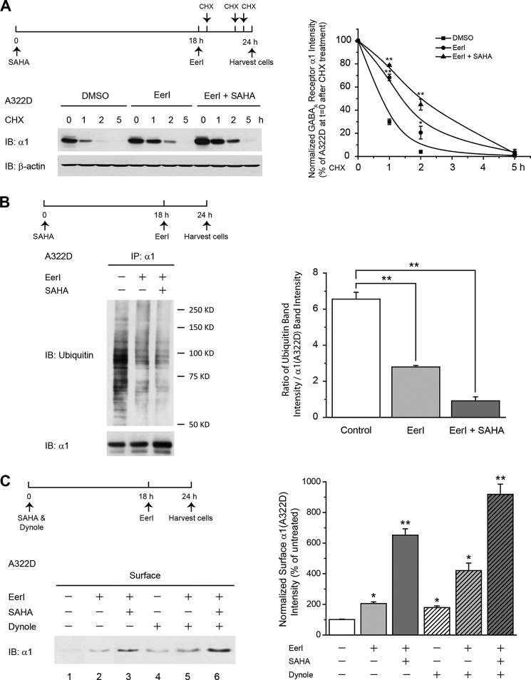
EerI treatment reduces the ERAD of the α1(A322D) subunit in HEK293 cells, and coapplication of SAHA further attenuates its ERAD without an apparent effect on its endocytosis. A, effect of EerI and SAHA on the degradation of the α1(A322D) subunit using CHX chase analysis. HEK293 cells stably expressing α1(A322D)β2γ2 receptors were treated with EerI (5 μm, 6 h) in the absence or presence of SAHA (2.5 μm, 24 h), followed by a chase with CHX (100 μg/ml), which inhibits protein synthesis, for the indicated time. Cells were harvested, lysed, and subjected to 8% SDS-PAGE gel and immunoblot (IB) analysis (n = 3). The dosage regimen is shown at the top. Degradation kinetics were plotted by quantifying α1(A322D) intensity against time after CHX addition. Data were normalized to the α1(A322D) intensity at t = 0 after CHX addition (right panel). DMSO, dimethyl sulfoxide. B, effect of EerI and SAHA on ubiquitinated α1(A322D) subunits. HEK293 cells stably expressing α1(A322D)β2γ2 receptors were treated with EerI (5 μm, 6 h) in the absence or presence of SAHA (2.5 μm, 24 h). Then the cells were lysed, immunoprecipitated using an anti-α1 antibody, and blotted for ubiquitin. The dosage regimen is shown at the top. The ubiquitin band intensity relative to α1(A322D) subunits post-immunoprecipitation (IP) was quantified and is shown in the right panel (n = 2). C, effect of EerI (5 μm, 6 h) and SAHA (2.5 μm, 24 h) on the cell surface α1(A322D) subunits in the absence or presence of a potent dynamin-1 inhibitor, dynole 34-2 (2.5 μm, 24 h). HEK293 cells stably expressing α1(A322D)β2γ2 receptors were treated with chemical compounds as in the dosage regimen. Then the surface α1(A322D) subunit was measured using a cell surface biotinylation assay. Quantification of the surface α1(A322D) subunit intensity is shown in the right panel (n = 3). Each data point in A, B, and C is reported as mean ± S.E. *, p < 0.05; **, p < 0.01.

