FIGURE 5.
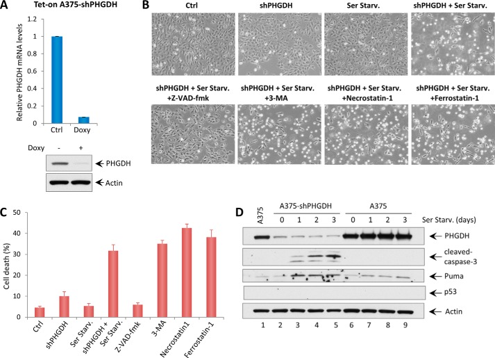
PHGDH knockdown induces apoptosis upon serine starvation. A, qRT-PCR analysis of PHGDH mRNA levels in the Tet-On A375-shPHGDH stable cell line after adding doxycycline (Doxy, 5 μg/ml) for 3 days. The Western blot analysis also shows the expression levels of PHGDH and Actin in Tet-On A375-shPHGDH cells after adding doxycycline. Ctrl, control. B, representative phase-contrast images of A375 cells expressing doxycycline-inducible shRNA against PHGDH with or without serine starvation (Ser Starv.) for 48 h. The images also show A375-shPHGDH cells under serine starvation conditions with the addition of cell death inhibitors (Z-VAD-fmk, a caspase 3 inhibitor, 10 μg/ml; 3-methyladenine (3-MA), an autophagy inhibitor, 2 mm; Necrostatin 1, a necroptosis inhibitor, 10 μg/ml; Ferrostatin 1, a ferroptosis inhibitor, 2 μm). C, the percentages of cell death for all experiments shown in B were measured by trypan blue exclusion assay. D, A375 shPHGDH cells and A375 cells were incubated in serine starvation medium for the indicated times, and total protein lysates were subjected to Western blot analysis for the expression of PHGDH, cleaved caspase 3, Puma, p53, and Actin.

