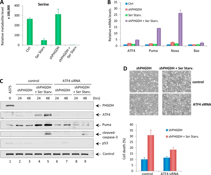
FIGURE 6.
ATF4 mediates PHGDH knockdown-induced apoptosis upon serine starvation. A, metabolite analysis showing the intracellular steady levels of serine in A375-shPHGDH cells cultured in complete medium or serine starvation medium (Ser Starv.). Ctrl, control. B, qRT-PCR analysis of mRNA levels of ATF4, Puma, Noxa, and Bax in A375-shPHGDH cells cultured in control medium or serine starvation medium. C, A375 shPHGDH cells were first transfected with control siRNA or ATF4 siRNA for 24 h and then incubated in complete medium or serine starvation medium for up to 48 h. Total cell lysates were harvested and subjected to Western blot analysis for the expression of PHGDH, ATF4, Puma, cleaved caspase 3, and p53. Vinculin was used as a loading control. D, representative phase-contrast images of A375-shPHGDH cells transfected with control or ATF4 siRNA in the presence or absence of serine at 48 h. The percentages of cell death were measured by trypan blue exclusion assay. Error bars represent mean ± S.D. from three experiments.

