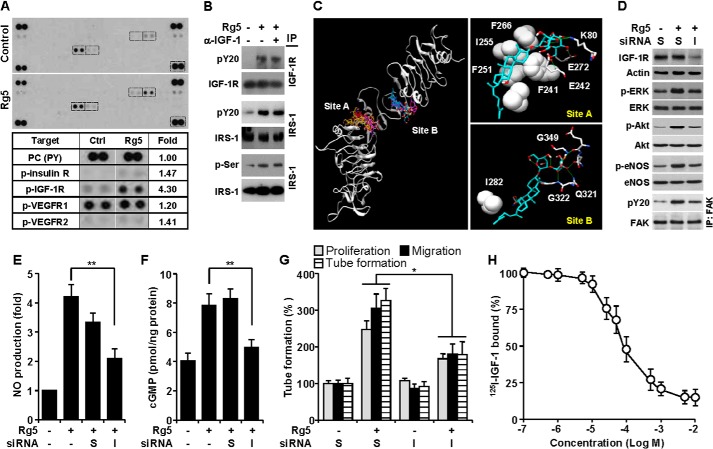
FIGURE 6.
Rg5-induced angiogenesis is mediated by IGF-1R activation. A, lysates of HUVECs stimulated with or without Rg5 (20 μm) for 30 min were applied to phospho-receptor tyrosine array kit. Phosphorylated receptors were determined, and the indicated boxes are compared in the lower panel. B, cells were stimulated with Rg5 (20 μm) alone or in combination with an anti-neutralizing IGF-1 antibody (α-IGF-1, 1 mg/ml), and levels of phospho-IGF-1R and IRS-1 were determined by immunoprecipitation (IP) and immunoblotting. C, in silico molecular docking analysis of Rg5 and IGF-1R was performed using Autodock 4.2 with a Lamarckian genetic algorithm. D–G, HUVECs were transfected with 100 nm scrambled (S) or IGF-1R siRNA (I) and stimulated with Rg5 (20 μm). D, after 30 min, the levels of target proteins were determined by immunoblotting. E, after 1 h, the intracellular levels of NO were determined by confocal laser microscopy using DAF-FM diacetate (n = 3). F, after 4 h, cellular cGMP levels were assessed by ELISA (n = 3). G, cell proliferation, migration, and tube formation were determined as described in the legend for Fig. 2 (n = 3). H, HUVECs were treated with 1 × 10⁻⁷-5 × 10⁻² m Rg5 for 20 min followed by incubation with 125I-labeled IGF-1 for 30 min. Cell-bound radiolabeled IGF-1 was counted by a scintillation counter (n = 3). *, p < 0.05, and **, p < 0.01.

