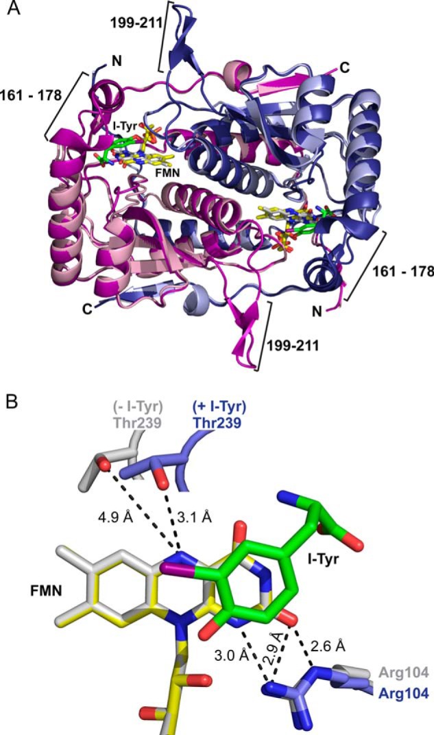
FIGURE 3.

Structure of hIYD. A, the structure of hIYD·I-Tyr (deep blue and deep purple to indicate the two monomers) is overlaid from a global fit to the structure of hIYD (light blue and light pink to indicate the two monomers). The active site lid composed of a loop (residues 199–211) and a helix-turn (residues 161–178) are only detected in the hIYD·I-Tyr co-crystal. I-Tyr is illustrated with its carbon skeleton in green. FMN is indicated in yellow and gray for structures containing and lacking the substrate I-Tyr, respectively. B, binding of I-Tyr to hIYD induces a reorientation of Thr-239 to provide a hydrogen bond to the N5 of FMN. This is illustrated with the same coloring used for the full structure in A and aligned relative to FMN.
