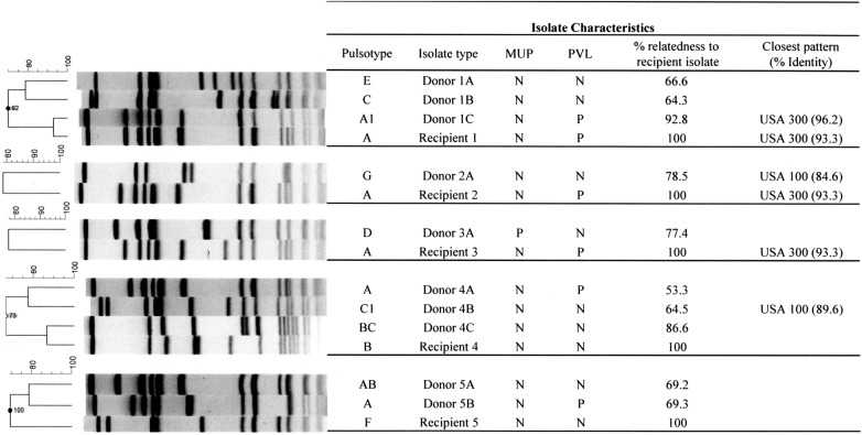
Figure 2.
Molecular characterization of “donor” and corresponding “recipient(s)” isolates. The pulsed-field gel electrophoresis (PFGE) dendogram compares fingerprint patterns of each recipient isolate (indicated by a letter) and their matching putative donor isolate(s) (indicated by the corresponding letter followed by numbers). Columns marked mupA and PVL (Panton-Valentine leukocidin) indicate the results for genetic tests performed to detect the mupA and PVL gene, respectively. Percentage relatedness to the recipient isolate and PFGE-type strains are indicated.

