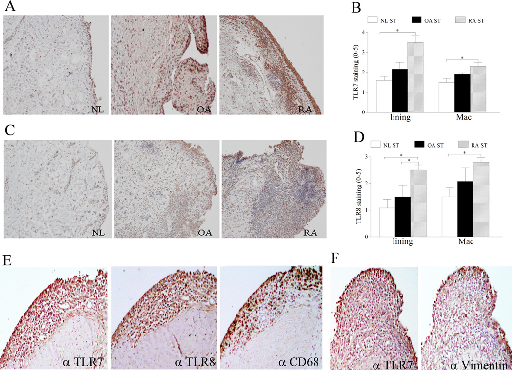
Figure 1. TLR7 and TLR8 expression is increased in RA synovial tissue (ST) lining and sublining macrophages compared to normal (NL) ST.
NL, OA and RA ST were stained with anti-human TLR7 (A) or anti-human TLR8 (C) (original magnification × 200) and positive immunostaining was scored on a 0–5 scale (B and D). ST lining and sublining macrophage immunostaining are shown as mean ±SEM, (n=5–7). E. RA serial sections were stained with anti-TLR7, anti-TLR8 and anti-CD68 antibodies in order to distinguish TLR7 and TLR8 staining on RA ST lining and sublining macrophages (original magnification × 400). F. RA serial sections were stained with anti-TLR7 and anti-vimentin antibodies in order to co-localize TLR7 staining in RA ST fibroblasts (original magnification × 400).

