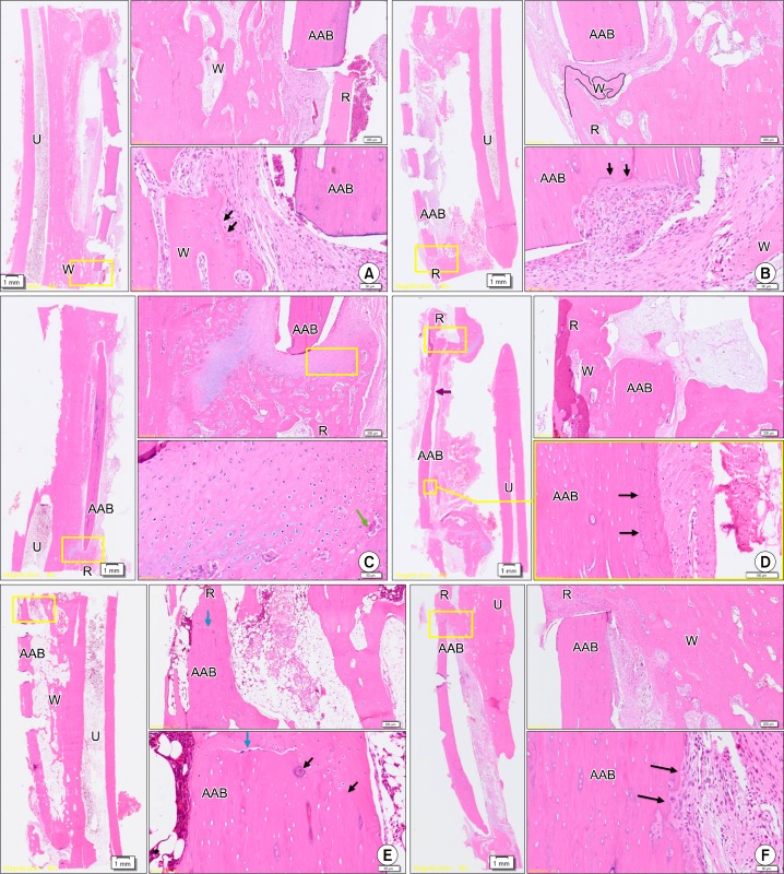
Fig. 6.
Histologic assessment of each animal at six and 12 weeks (A∼C: six weeks after surgery, D∼F: twelve weeks after surgery). (A) Group 1, six weeks after surgery. Although woven bone originated from the adjacent ulnar bone and a reversal line, suggesting bony remodeling, was observed, the new bone did not unite with the AAB. (B) Group 3, six weeks after surgery. Although woven bone originated from the adjacent ulnar bone and a reversal line which suggests bony remodeling was observed, the new bone did not unite with the AAB. (C) Group 2, six weeks after surgery. High magnification revealed extensive cartilage formation around the AAB, active cell division of chondroblasts, and replacement of cartilage by osteoid. (D) Group 1, twelve weeks after surgery. Some osteoclasts and osteoblasts were also observed, suggestive of bony remodeling, and woven bone originated from a single end of the radial bone and restored some continuity with the AAB. Sparse new bone formation and a reversal line suggested proper engraftment of AAB. (E) Group 2, 12 weeks after surgery. Woven bone originated from the adjacent cutting ends of the radius and showed complete fusion with AAB. At low magnification, AAB had completely restored continuity with the radial bone marrow. Moreover, high magnification revealed reversal lines and osteocytes inside the woven bone, suggesting that active bony remodeling was in progress. (F) Group 3, twelve weeks after surgery. There was some evidence suggesting bony remodeling around AAB, similar to the group 1 results, although not as extensive as in group 2 (H&E; left: ×40, right: ×100/×400). Boxes with yellow line in left slides (×40) were magnified in right slides (×100/×400) to be determined whether complete bony fusion with osteoblast, osteoclast, and osteoid was done. U, ulnar bone; R, radial bone; AAB, autoclaved autogenous bone; W, woven bone; black arrows, reversal line; blue arrows, end of AAB; purple arrows, resorption of AAB; green arrow, osteoclast.

