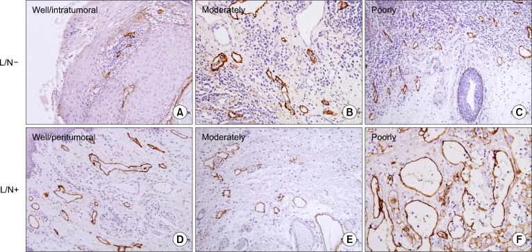
Fig. 2.
Lymphatic vessel density (LVD) and lymphatic vessel dilatation; structures stained with D2–40 (brown) are compressed, apparently non-functional lymphatics (A: intratumoral LVD). Functional lymphatics are dilated and have larger surface area (D–F: peritumoral LVD), higher number in lymph node+ (L/N+) group. We can see clearly the unstained microvessels (A∼D). Upper panel is L/N− group and lower panel is L/N+ group. Representative slides are shown (A∼F: ×200) (counterstained by Mayer’s hematoxyline).

