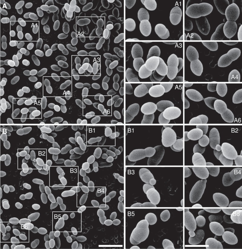
FIG 2 .
Scanning electron microscopy of ΔlocZ mutant (Sp57) and wild-type S. pneumoniae (Sp1) cells. (A and B) Overviews of the WT (A) and the ΔlocZ mutant (B). The boxes indicate the areas shown in the panels on the right. In panels A1 to A6, WT cells with the characteristic diplococcal morphology, including individual cells (A5) or cells at different stages of cell division (A1 to A4 and A6), can be seen. Dividing cells are similar in shape and size, with septa perpendicular to the long cell axis. In panels B1 to B6, altered morphology of the ΔlocZ mutant is presented. Panel B1 shows that Sp57 dividing cells are uneven in shape and size. The cell poles are deviated from the long cell axis. A typical potato-like cell is shown in the upper right corner. In panels B2 to B4 and B6, typical minicells can be seen at one of the cell poles. B5 shows an unusual cell with two septa not perpendicular to the long cell axis. The magnifications of the images in panels A and B are the same. Bar, 2 µm. The images in panels on the right are magnified twice. Bar, 1 µm.

