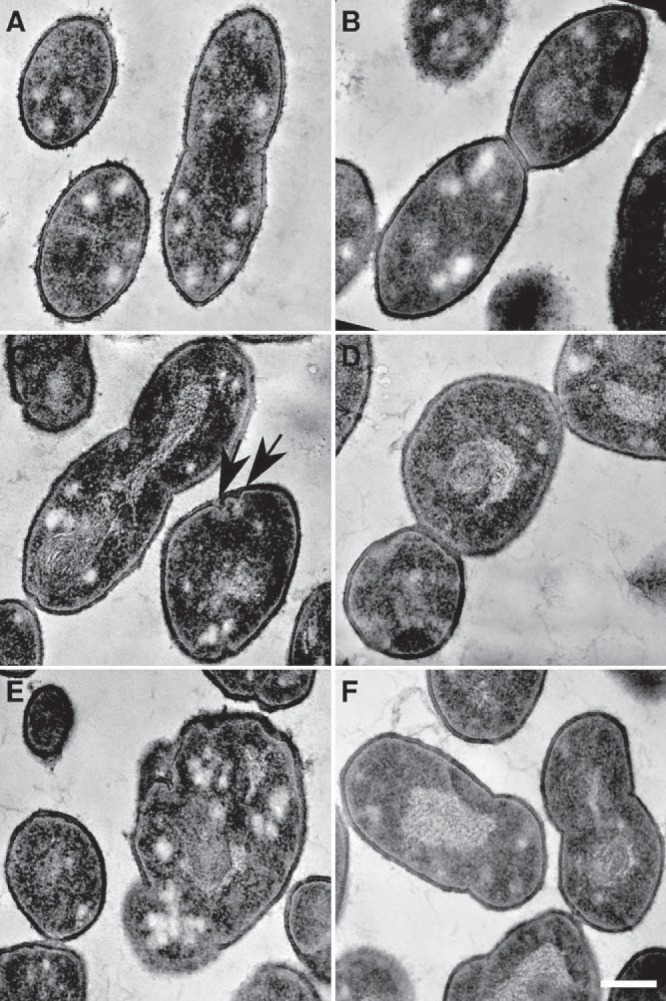
FIG 3 .

Transmission electron microscopy of ΔlocZ mutant (Sp57) and wild-type S. pneumoniae (Sp1) cells. (A and B) Typical cell division of WT strain. (A) Beginning of septum formation. (B) Cells with well-developed septum, just before their separation. (C to F) Altered ultrastructure of the ΔlocZ mutant. (C) Example of the configuration of several septa; invaginations in close proximity are marked by arrows. (D) Example of three cells still connected by the septa. (E) Example of bacterial cell lysis; the cell wall rupture and altered cytoplasm can be seen. (F) Uneven cell division resembling the cells shown in Fig. 2B, subpanel B1. Magnifications are the same for all images. Bar, 0.2 µm.
