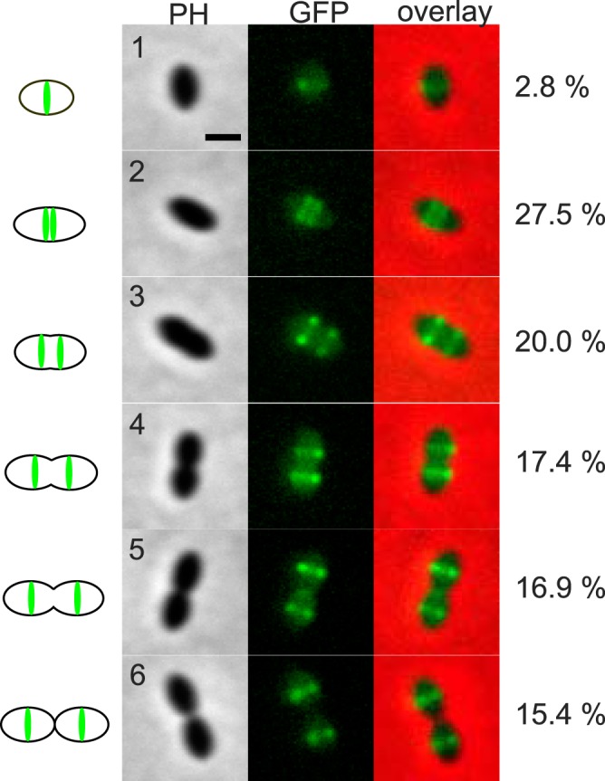
FIG 4 .

Localization of GFP-LocZ in S. pneumoniae. A representative cell for each conventional stage of the pneumococcal cell cycle (designated 1 to 6) illustrates the localization of GFP-LocZ expressed under the control of its native promoter (strain Sp229). Phase contrast (PH), GFP signal (GFP), and overlays are shown. Schematic pictures on the left document the position of the GFP signal in the cells. The percentages of cells at each stage over 610 cells are shown on the right. All cells at each stage showed the same localization profile as did the representative cell shown in the figure. Bar, 1 µm.
