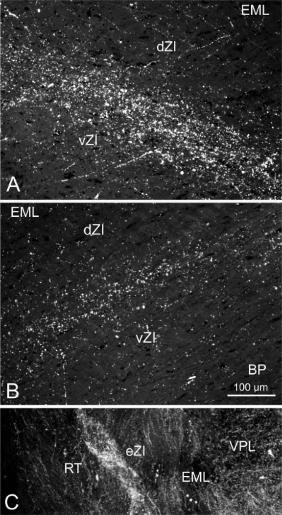
Fig. 3.
Photomicrographs of corticoincertal terminals labeled from injections of WGA-HRP into cortex, as observed with crossed polarizing filters. A: Labeled terminals located in vZI are densest along the border between the sublaminae following an injection of the SI cortex face representation. B: The pattern of terminal label in ZI forms a band of label located at the border between the sublaminae following a WGA-HRP injection in the FEF. C: Terminal patches were interspersed between bundles of labeled fibers in eZI in this example from an injection of the SI cortex forepaw representation.

