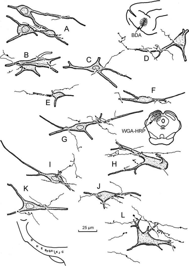
Fig. 5.
High-magnification illustration showing the spatial relationship between BDA-labeled corticoincertal terminals from an SI cortex injection (insert upper right) and WGA-HRP-labeled neurons within ZI from an injection in the SC (insert middle right). The location of the illustrated elements is shown schematically at the lower left.

