
Figure 1.
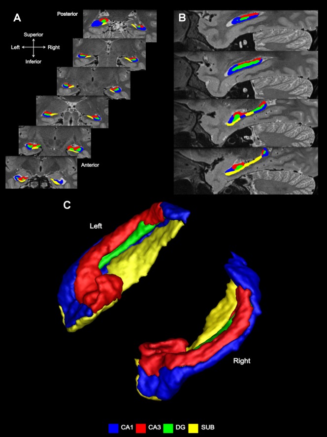
Subfield segmentation. (A) In the coronal plane—coronal sections through an averaged T2-weighted image of the left and right hippocampus of an example participant. (B) Subfield segmentation in the sagittal plane. (C) An example of subfield segmentation in 3D. [Color figure can be viewed in the online issue, which is available at http://wileyonlinelibrary.com.]