Figure 1.
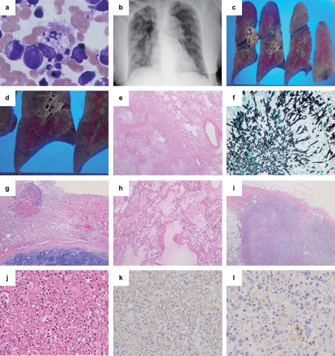
Clinical images and pathological findings of Case 1. (a) Bone marrow finding and (b) chest X-ray image, (c,d) gross findings in the lungs, (e,g–j) hematoxylin and eosin staining, (f) Grocott staining, and (k,l) immunohistochemistry (IHC) using anti-SFTSV-NP antibody. (a) In the bone aspirate, many histiocytes show hemophagocytosis (×400). (b) A chest X-ray reveals a bilateral infiltrative shadow without consolidation. The cut surface of the right lung shows (c) many dispersed white nodular legions, (d) mainly in the lower lobe. (e) In the lung, there is necrotizing inflammation (×40) and (f)Aspergillus infection in the nodular lesions (Grocott staining ×400). (g) A tracheal ulcer with Aspergillus was also noted (×100). (h) Hyaline membrane formation indicating secondary diffuse alveolar damage is seen (×100). (i,j) In the left inguinal lymph node, the basic architecture of the lymph node is replaced by massive necrosis with infiltration of lymphocytes, histiocytes, some atypical lymphoid cells, and a significant amount of nuclear debris, but no neutrophils are observed (i, ×40; j, ×200). (k) In IHC of the lymph node, SFTSV-NP-positive cells are found (×100), and (l) positive staining for the SFTSV-NP antigen is detected in the cytoplasm of atypical lymphoid cells (×400).

