Figure 2.
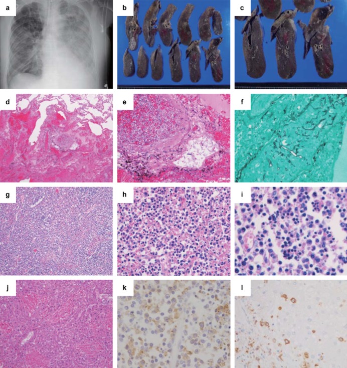
Clinical images and pathological findings of case 2. (a) Chest X-ray image, (b,c) gross findings in the lungs, (d,e,g–j) hematoxylin and eosin staining, (f) Grocott staining, and (k,l) immunohistochemistry (IHC) using anti-SFTSV-NP antibody. (a) A chest X-ray reveals a bilateral infiltrative shadow without consolidation. (b,c) The cut surface of the right lung shows foci of pulmonary hemorrhage and infarction. (d) Diffuse hemorrhagic infarction (×40) and (e,f) angio-invasion of Mucor (e, ×200; f, Grocott staining ×400) are seen in the lung. (g,h) Necrotizing lymphadenitis is present in the lymph node around the abdominal aorta (g, ×40; h, ×400), and (i) hemophagocytosis is also observed (i, ×400). (j) The liver shows lobular necroses and mild portal fibrosis (×100). (k) IHC of the lymph node (×400) shows numerous SFTSV-NP-positive cells and positive signals for the SFTSV-NP antigen in the cytoplasm of atypical lymphoid cells. (l) IHC of the liver shows SFTSV-positive cells, but hepatocytes were negative for the SFTSV-NP antigen (×100).

