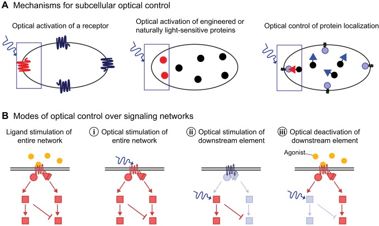
Fig. 2.
Modes of optical control. (A) Local photoactivation of a naturally light-sensitive receptor can be used to trigger signaling from a selected region at the cell surface (shown on the left). Inside the cell, signaling can be locally triggered by optical activation of naturally light-sensitive adenylyl cyclases or engineered fusions of signaling proteins with light-sensitive domains that mask or unmask their active sites in response to photoactivation (middle). Photoactivation of selected protein domains (triangles) can be used to translocate signaling proteins to specific subcellular sites, such as the plasma membrane at one side of the cell (right). (B) Optical control can be used to activate or inhibit signaling at different nodes within a signaling cascade. Optical activation of a receptor stimulates the entire signaling network (i). Alternatively, a downstream signaling protein can be directly activated independently of any receptor activity upstream (ii). Finally, a receptor is activated through its ligand, but a downstream protein is inhibited optically (iii). Activated proteins are in red and inactive proteins are in light blue. Filled blue circles are receptor-activating ligands. Wavy arrow denotes light. Activated components are shown in red and inactive in blue and black.

