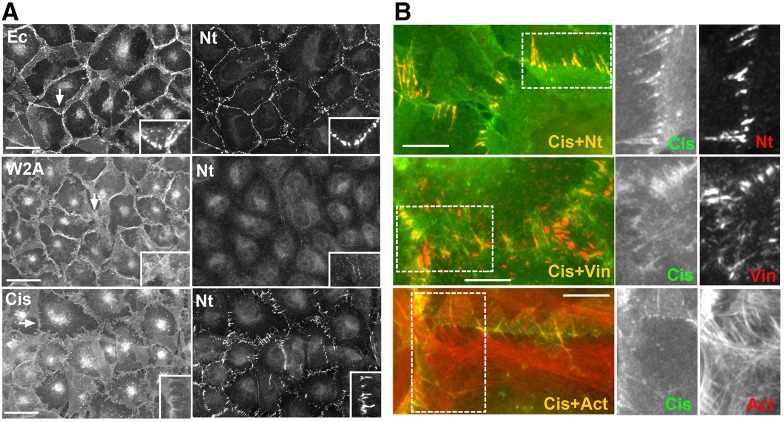
Fig. 2.
Nectin-2 localization depends on cadherin adhesion properties. (A) A431D cells stably transfected to express EcadDn (Ec) or its mutants bearing point mutations inactivating either its adhesive (W2A) or cis-binding (Cis) interfaces. Cells were double-stained for cadherin (left column) and nectin-2 (Nt, right column). Higher magnifications of the selected regions (indicated by arrows) are shown in the insets. Scale bars: 30 µm. (B) High magnifications of the junctions produced in A431D cells expressing the cis-EcadDn mutant. The cells were double-stained for the mutant (Cis, green) and for nectin-2 (Nt), vinculin (Vin) or actin (Act). Green and red channels are shown separately for the boxed regions. Note that the cells contact one another through numerous actin-rich filopodia co-recruiting nectin and vinculin. Scale bars: 10 µm.

