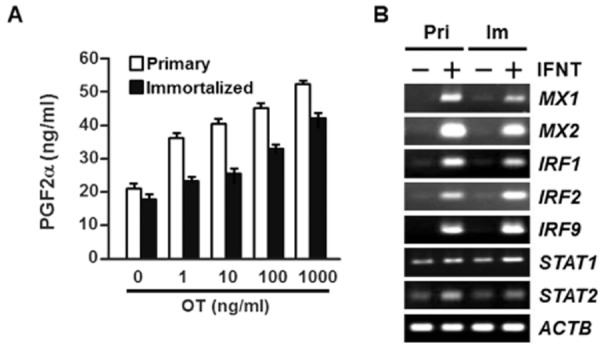
Figure 3.

Effect of OT on PGF2α accumulation and the expression of interferon (IFN) stimulated genes following recombinant ovine IFNT treatment in immortalized endometrial epithelial cells (imEECs). (A) The primary (white bar) and immortalized at 60th passage (black bar) EECs were cultured on 48-well plates coated with collagen type IA. The cells were treated with increasing concentrations of OT (0, 1, 10, 100 or 1000 ng/mL) and PGF2α accumulation in conditioned media after 24 h incubation were measured using an enzyme immunoassay. The concentrations were corrected for cell numbers as determined by Cell Counting Kit-8 (CCK-8) (see Materials and Methods). The mean concentration of prostaglandin F2α (PGF2α) was derived from an average of three wells within a single plate experiment. Values represent means ± SEM. (B) The expression of IFN-stimulated gene transcripts in primary and immortalized EECs. The primary (Pri) and immortalized (Im) EECs were seeded on six-well plates coated with collagen type IA. The cells were treated with IFNT (1 μg/mL) for 24 h and the expression of IFN stimulated gene transcripts was analyzed by RT-PCR. IFNT stimulated genes include interferon-inducible Mx proteins (Mx1 and Mx2), interferon regulatory factors (IRF1, 2 and 9) and signal transducers and activator of transcriptions (STAT1 and 2). ACTB was used as an internal control.
