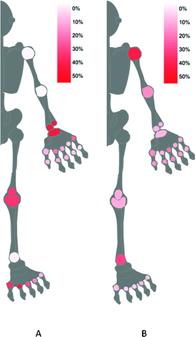
Figure 1.

Graphical summary (homunculus) of A, subclinical synovitis (shown in red), graded according to the proportion of joints in which ultrasound (US) was positive (gray-scale [GS] score ≥2 or power Doppler [PD] score ≥1 present) and clinical examination (CE) was negative (no tenderness or swelling) and B, apparent clinical overestimation of synovitis (shown in red), graded according to the proportion of joints in which US was negative (GS score ≥2 or PD score ≥1 absent) and CE was positive (tenderness and/or swelling present). White = 0% of joints; red = 50% of joints.
