
Figure 1. The Eya1+ IM Contributes to Caudal Mesonephric and Metanephric Nephrons.
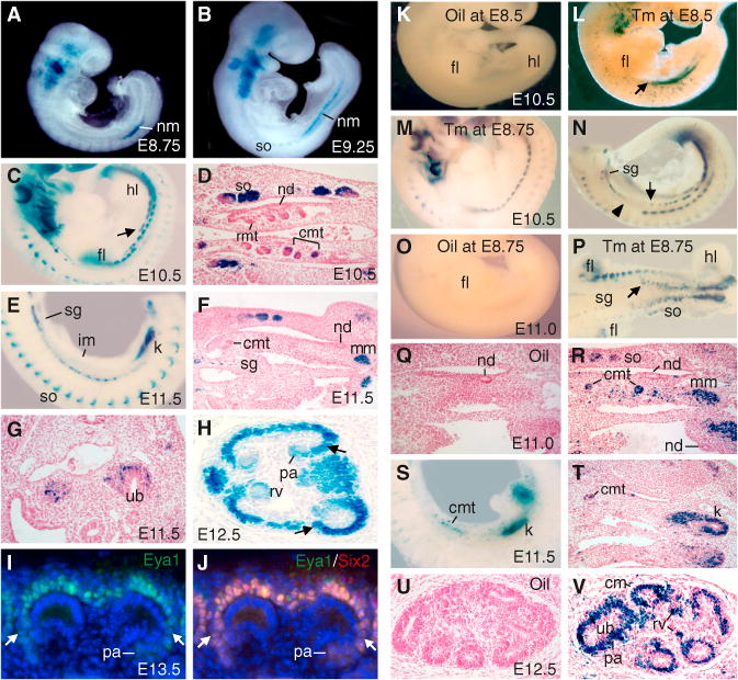
(A–C and E) β-gal staining of an Eya1LacZ/+ embryo at E8.75 (A), E9.25 (B), E10.5 (C), and E11.5 (E). The arrow points to the Eya1+ IM.
(D and F) Sections of β-gal+ embryos at E10.5 (D) and E11.5 (F).
(G and H) β-gal staining on E11.5 (shorter staining) (G) and E12.5 (H) kidney sections. The arrows in (H) point to weaker activity of the LacZ+ subregion. (I and J) Immunostaining for Eya1/Six2on anE13.5 kidney section, showing Eya1 alone (I) and a merged image for both Eya1 (green) and Six1 (red) (J). The arrows point to lower levels of the Eya1/Six2 subregion.
(K–V) Fate mapping of Eya1+ cells in Eya1CreERT2/+; R26RLacZ/+ embryos at E10.5–E12.5 after injection of oil (K, O, Q, and U) or 2–3 mg Tm (L–N, P, R–T, and V) at E8.5–E8.75. (K–O and S) show whole-mount lateral or (P) ventral views. The arrows point to the anterior limit of the Eya1+ IM. The arrowhead points to the forelimb region. (Q, R, and T–V) show sections counterstained with hematoxylin.
cmt, caudal mesonephric tubule; fl, forelimb; hl, hindlimb; k, kidney; nd, nephric duct; nm, nephrogenic mesoderm; rmt, rostral mesonephric tubule; sg, sympathetic ganglion; so, somite. See also Figure S1.