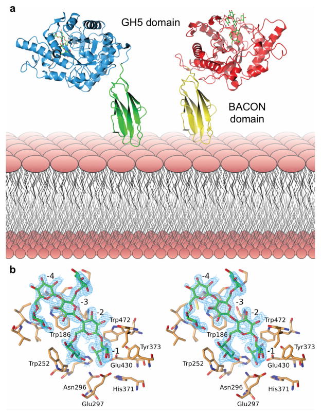
Figure 4.
Structural biology of BoGH5A. a. Tertiary structure; the two conformations observed in crystallo have been oriented relative to the N-terminal, membrane-anchored BACON domain (see also Supplementary Video V1). b. Wall-eyed stereo view of the binding of XXXG in the -4 to -1 subsites (see also Supplementary Video V2). The wireframe represents an unbiased 2Fo-Fc map (contoured at 0.3 electrons per Å3) obtained using phases calculated from the best model prior to the incorporation of any ligand in refinement.

