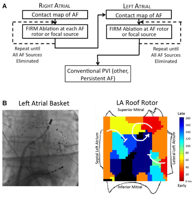
Figure 1.
(A) Workflow for FIRM-guided ablation of AF. (B) Typical basket placement and results from FIRM-guided ablation. Basket placed in left atrium, with resulting FIRM map showing AF rotor at roof with surrounding spiral arms disorganizing and fusing with the fibrillatory milieu (blocked arrows) and rotor precession within a limited area on successive cycles (not shown).

