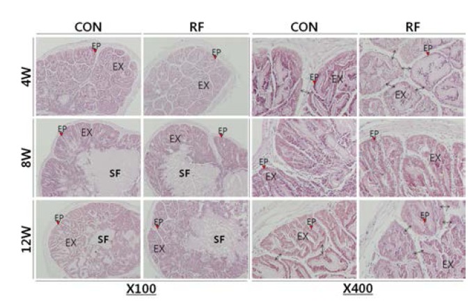
Fig. 2.

Histological analysis of mice seminal vesicle. Seminal vesicles from night feeding (CON, control, 17:00-21:00) and reverse feeding (RF; day feeding, 09:00-13:00) mice were fixed. The specimens cut at 6 μm, then the sections were stained with hematoxylin-eosin (H.E.) stain and examined under light microscope. EP, epithelial cell; EX, papilla folding; SF, seminal fluid.
