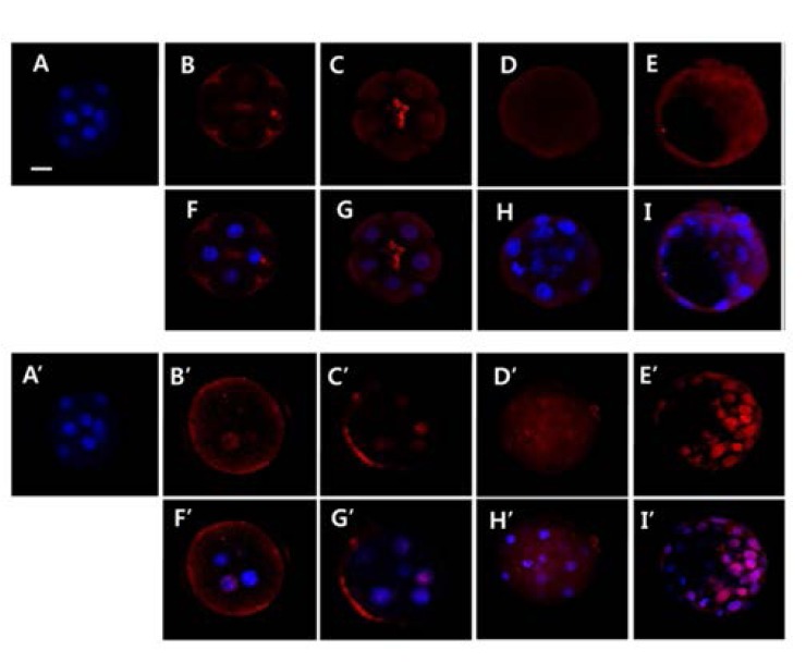
Fig. 3.

Laser scanning confocal microscopy image of AQP5 in vivo (A-I) and in vitro (A’-I’) developed embryos. The localization of AQP5 in embryo was examined by whole mount immunofluorescence. A and A’: negative control; B and B’: 4-cell stage embryo; C and C’: 8-cell stage embryo; D and D’: morula stage emryo; E and E’: blastocyst stage embryo; AD and A’-D’: red (anti-AQP5); F-I and F’-I’: merged image (blue: Hochest33238); A-I: in vivo embryos; A’-I’: in vitro cultured embryos from 2-cell stage; scale bar = 20 μm.
