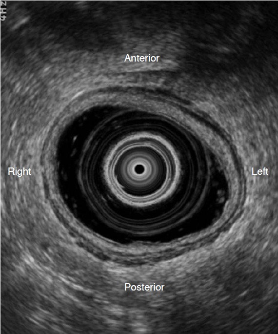
Fig. 1. Optimal transrectal ultrasonography scan.

The axial view clearly shows the layers of the rectal wall with proper rectal distension after cleansing with an enema. The transducer is placed in the center of the rectal lumen.

The axial view clearly shows the layers of the rectal wall with proper rectal distension after cleansing with an enema. The transducer is placed in the center of the rectal lumen.