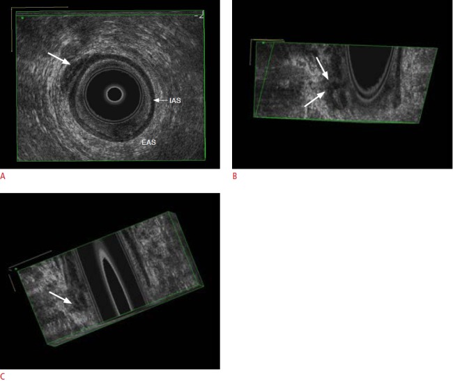
Fig. 10. Intersphincteric perianal fistula.

A. The hypoechoic tract (arrow) is seen between the internal (IAS) and external anal sphincters (EAS) in the axial image. B, C. Threedimensional reconstructed sagittal (B) and coronal (C) images better represent the exact location of the fistula (arrows).
