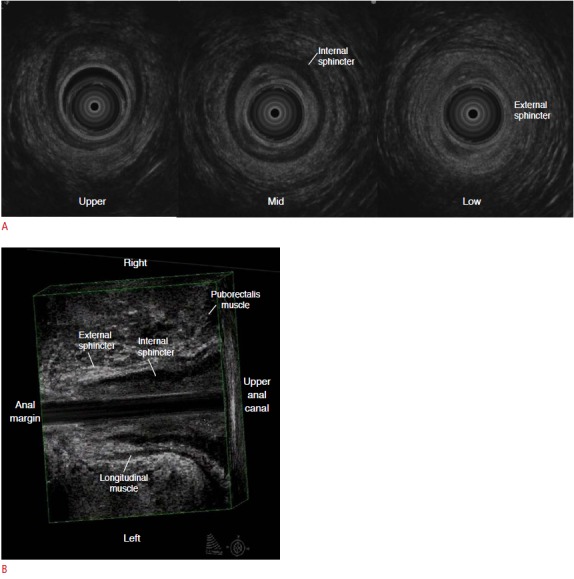
Fig. 4. Normal ultrasonogram of the anal canal.

A. The anal canal is usually divided into three levels for examination. In the upper anal canal, the puborectalis muscle is seen as a U-shaped echogenic band. In the middle anal canal, the internal anal sphincter is most clearly seen as a thickened hypoechoic layer. In the lower anal canal, the echogenic external anal sphincter is seen together with the termination of the internal anal sphincter. B. On coronal reformatted three-dimensional ultrasonogram, a hypoechoic longitudinal layer indicates the internal sphincter, terminating at the lower anal canal. The external anal sphincter is represented by a hyperechoic layer running through the outer aspect of the internal anal sphincter.
