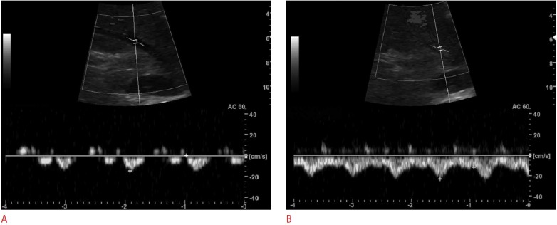
Fig. 2. Doppler wave form change of middle hepatic vein to biphasic pattern with elevated baseline during the Valsalva maneuver.

A. At rest, biphasic pattern is seen in a subject. B. During Valsalva maneuver, baseline is elevated and peak velocity is maintained. This peak velocity was thought as a dominant antegrade wave caused by suction of blood into the right atrium from the liver due to the tricuspid annulus which moves toward the cardiac apex during ventricular systole.
