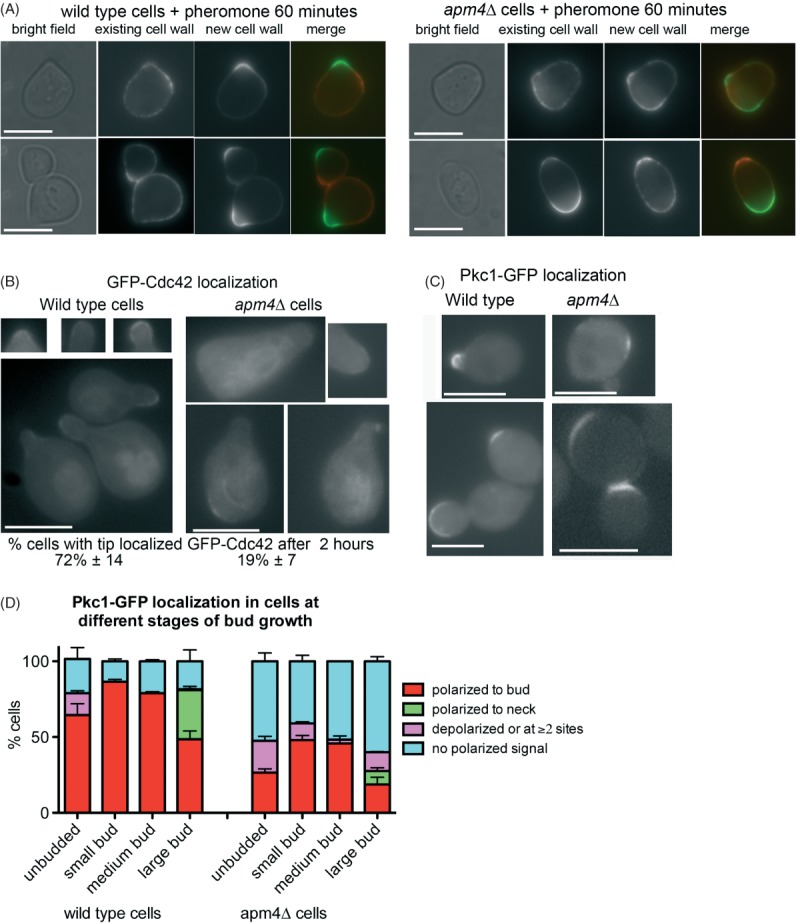
Figure 3.

Defects in polarity of cell wall markers in apm4Δ cells. A) Cells were incubated with concanavalin A conjugated to distinct fluorophores to label old and new growth of cells. Left panels show representative wild type cells; right panels show labelled apm4 null cells. Scale bar = 5 µm. B) Wild type and apm4Δ cells were induced to express GFP-Cdc42 by growth in galactose containing medium for 3 h before addition of alpha factor for 2 h. Localization of GFP-Cdc42 was analysed in 100 cells in three independent experiments. Scale bar = 5 µm. C) Pkc1-GFP was analysed in cells to determine localization under conditions of vegetative growth. Scale bar = 5 µm. Shown are cells from different stages of budding. D) Localization of Pkc1-GFP was analysed in cells and extent of polarity recorded and shown graphically. Data are from three independent experiments with >100 cells counted in each experiment.
