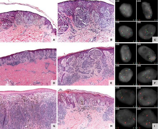
Figure 1.
Spitz/Spitzoid lesions. (A–C) Specimen from a 10-year-old boy with a 4-mm brown papule on the left upper arm. (A, B) Spitz nevus—small, well-circumscribed, symmetric junctional melanocytic lesion composed of vertically oriented nests of spindled to epithelioid melanocytes with abundant cytoplasm, large nuclei, prominent nucleoli, and prominent pagetoid scatter of the melanocytes. (C) Fluorescence in situ hybridization (FISH) negative. (D–F) Specimen from a 25-year-old woman with a right lower leg lesion. (D, E) Atypical Spitz tumor. Compound melanocytic proliferation, with spindle features compatible with a Spitz nevus. Cytologic features vary from larger discohesive cells with prominent nucleoli to smaller mature melanocytes in the dermal component. Mild inflammatory infiltrate associated with this lesion and occasional epidermal Kamino bodies. No significant pagetoid spread or dermal mitotic figures. Hyperchromasia of dermal melanocytes, dyshesion within epidermal nests, and lack of definitive dermal architectural and cytologic maturation are seen. (F) FISH negative. (G–I) Specimen from a 16-year-old boy with a dark brown macule with central black papule, slowly enlarging. (G, H) Spitzoid melanoma. Atypical but well-defined compound melanocytic lesion, asymmetric with heavy, brisk lymphocytic host response and uneven pigmentation. Prominent, multifocal pagetoid upward scatter of malignant melanocytes into the epidermis. The tumor expands the dermis and focally consumes the epidermis. The atypical melanocytes are epithelioid with pink–brown cytoplasm and pleomorphic, irregular nuclear contours. Frequent mitotic figures are identified and enumerated at 3/mm2. (I) FISH positive (RREB and CCND1 positive) of uncertain malignant potential.

