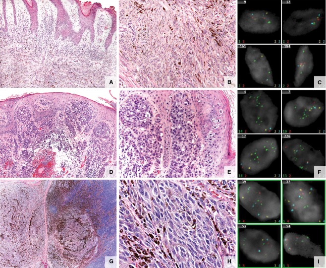
Figure 2.
Atypical melanocytic lesions. (A–C) Specimen from a 20 year-old man with a left posterior scalp lesion. (A, B) Atypical melanocytic tumor of uncertain malignant potential (MelTUMP). Infiltrative predominantly intradermal spindle cell melanocytic proliferation extending to subcutaneous tissue. The epidermis is irregularly acanthotic without evidence of ulceration. Composed of spindled melanocytes in intersecting fascicles. A dermal mitotic figure is identified on hematoxylin and eosin stained sections. Melan-A and Ki-67 double-labeled immunohistochemical stain failed to identify a proliferating dermal melanocytic population. (C) Fluorescence in situ hybridization (FISH) negative. (D–F) Specimen from a 16-year-old girl with scalp melanoma. (D, E) Melanoma. Extensive epidermal ulceration and a markedly atypical compound melanocytic population of lentiginous, haphazardly nested spindled melanocytes with dusky cytoplasm, pleomorphic nuclei, and prominent nucleoli within the epidermis with extensive pagetoid scatter. Kamino bodies are not appreciated. Atypical cells extending from the epidermis through the dermis into the subcutaneous fat demonstrate numerous mitotic figures (5/mm2) and lack maturation with downward descent. There is ulceration and prominent epidermal consumption by the melanocytic proliferation. Focally, areas suggestive of regression are noted at the epidermal surface of the lesion. (F) FISH positive (CCND1 positive). (G–I) Specimen from a 15-year-old boy with an enlarged mastoid lymph node. The patient had an initial atypical compound melanocytic proliferation of the right scalp diagnosed at 13 years of age. (G, H) Metastatic melanoma. Lymph node extensively infiltrated by an array of atypical pigmented cells with high nuclear to cytoplasmic ratios, irregular nucleoli, and dense chromatin. Heavy pigmentation is present in some of the cells. Lesional cells are mitotically active and zones of necrosis and areas of fibrosis are seen. (I) FISH positive (RREB and CCND1 positive).

