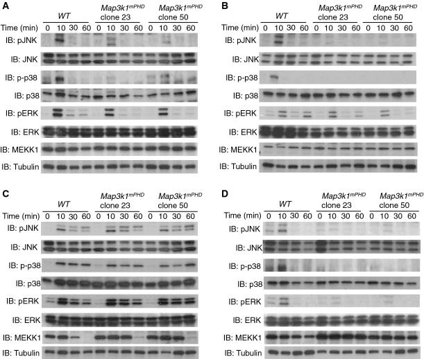
Figure 2. Map3k1mPHD ES cells exhibit defective JNK and p38 activation following TGF-β, EGF and nocodazole stimulation.
A–D WT and Map3k1mPHD ES cells were kept on low serum and stimulated with (A) TGF-β (10 ng/ml), (B) EGF (100 ng/ml), (C) sorbitol (500 mM) or (D) nocodazole (0.5 μg/ml) for 10, 30 and 60 min or left unstimulated. Cells were lysed and analysed by IB using the indicated antibodies.
Data information: Results are representative of three independent experiments.
Source data are available online for this figure.

