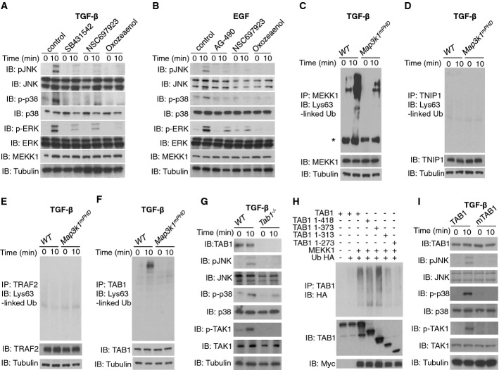
Figure 4. MEKK1 PHD dependence of TGF-β-stimulated TAK1 and MAPK signalling.
A WT ES cells were rested in low serum conditions and stimulated for 10 min with TGF-β (10 ng/ml) in the presence or absence of DMSO (control), SB431542, NSC697923, (5Z)-7-Oxozeaenol (Oxozeaenol) or left unstimulated. Lysates were made and analysed by IB using the indicated antibodies.
B WT ES cells were rested in low serum and stimulated for 10 min with EGF in the presence or absence of DMSO (control), AG-490, NSC697923, (5Z)-7-Oxozeaenol (Oxozeaenol) or left unstimulated. Lysates were prepared and analysed as above.
C–F WT or Map3k1mPHD ES cells kept in low serum were stimulated or not for 10 min with TGF-β (10 ng/ml). Lysates were prepared and IP and IB performed using the indicated antibodies (* indicates a non-specific band).
G WT or Tab1−/− ES cells were analysed as above by the indicated antibodies before and after TGF-β (10 ng/ml) stimulation.
H HEK 293 cells were transfected with the indicated constructs and lysates made. IP and IB were performed using the indicated antibodies.
I Tab1−/− ES cells were transfected with TAB1 or mTAB1 as indicated, rested in low serum and stimulated or not for 10 min with TGF-β (10 ng/ml). Lysates were made and analysed as above.
Data information: Results are representative of three experiments.
Source data are available online for this figure.

