
Figure 1.
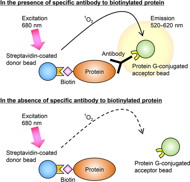
Schematic illustration of the AlphaScreen method for detecting a biotinylated protein–antibody interaction. The streptavidin-coated donor beads interact with the biotinylated target protein. The protein G-coated acceptor beads interact with the target protein via the specific antibodies. When the 680 nm excitation light is given, the donor beads generate singlet oxygens. The singlet oxygens promote the acceptor beads to emit luminescent light at 520–620 nm. This reaction occurs only in the presence of the specific antibodies in the solution, since the antigen–antibody reaction shortens the distance between both beads within 200 nm (top panel). No luminescence is seen without specific antibodies (bottom panel).