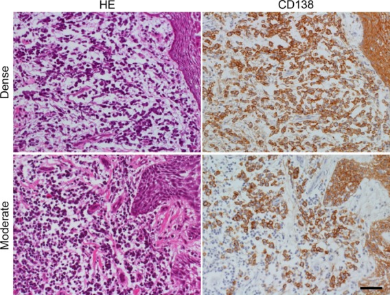
Figure 3.

Plasma cells in consecutive frozen sections of the biopsied gingiva. Two representative lesions of gingivitis demonstrate dense (top panels: case 1) and moderate (bottom panels: case 9) infiltration of plasma cells beneath the squamous lining. Left panels: hematoxylin & eosin staining, right panels: immunostaining for CD138. Not only plasma cells located in the subepithelial layer but also covering squamous epithelial cells reveal membrane positivity for CD138. Bar indicates 50 μm.
