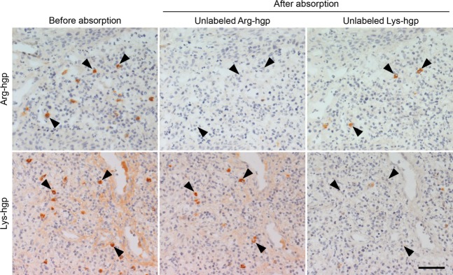
Figure 6.

Absorption experiment using unlabeled Arg-hgp and Lys-hgp. The enzyme-labeled antigen method for Arg-hgp reactivity and Lys-hgp reactivity on consecutive frozen sections in case 1 is illustrated. The reactivities to Arg-hgp and Lys-hgp in the cytoplasm of plasma cells (left panels) are abolished with an excess amount of the corresponding unlabeled proteins (center top and right bottom panels), and partly eliminated with unlabeled Lys-hgp and Arg-hgp, respectively (center bottom and right top panels). Arrowheads indicate plasma cells producing antibodies reactive to the unique epitope on Arg-hgp or Lys-hgp. Bar indicates 50 μm.
