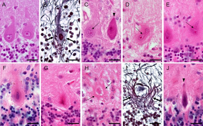
Figure 1.
Changes of Purkinje cell morphology in cases with spinocerebellar ataxia type 31 (SCA31) (A and B: control subject; C–J: SCA31 case). (A) Purkinje cells of a control subject have round nuclei. (B) Thin neurites (arrows) around a Purkinje cell are seen in a control subject. (C) Two normal-sized Purkinje cells are shown in an SCA31 case. One has a dark cytoplasm and nucleus, suggesting that it has started to shrink (arrowhead). The other apparently has the halo-like amorphous materials with a bent nucleus (thin arrow). (D and E) Bright and elongated nuclei (thin arrows) are occasionally folded up at their end (D, thin arrow) in Purkinje cells with the halo-like amorphous materials. (F) Degenerated Purkinje cell with the typical halo-like amorphous materials. Purkinje cells are shrunken and their nuclei become severely deformed, rod-shaped, and condensed. (G) Ghost-like feature of a Purkinje cell is shown, where the outline of the nucleus is obscure. (H) Ghost-like feature of a Purkinje cell surrounded by bundles of lines (thin arrows). (I) The cell body of Purkinje cell is obscured in the empty basket formed by thick neurites (arrows). (J) A severely thin Purkinje cell with a shrunken and condensed nucleus that was not surrounded by the halo-like amorphous materials. A, C–H, and J: HE stain, B and I: Bodian stain. Bars; 20 μm.

