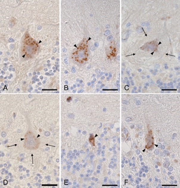
Figure 4.

Immunohistochemistry for trans-Golgi network protein 2 (TGOLN2). (A) Granular appearance of a Purkinje cell from a control subject (arrowheads). (B) Normal-sized Purkinje cells without the halo-like amorphous materials from case 1 with spinocerebellar ataxia type 31 (SCA31) show the relative preservation of the Golgi apparatus (arrowheads). (C and D) Purkinje cells with the halo-like amorphous materials (arrows) from case 1 (C) and 2 (D) with SCA31 show severely fragmented and a reduced amount of the Golgi apparatus (arrowheads). (E and F) Shrunken Purkinje cells without the halo-like amorphous materials from cases 1 (E) and 2 (F) with SCA31 show reduced, but not fragmented Golgi apparatus (arrowheads). Bars; 20 μm.
