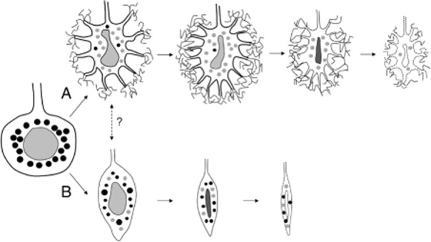
Figure 5.

Schematic drawing of the chronological changes of Purkinje cell morphology in spinocerebellar ataxia type 31 (SCA31). Two different processes of Purkinje cell degeneration are shown. One is the shrinkage of Purkinje cells with the halo-like amorphous materials (A). These cells very often have bent and elongated nuclei. The other is the shrinkage of Purkinje cells without the halo-like amorphous materials (B). These cells develop marked atrophy with slender and condensed nuclei. The black circles in the cytoplasm indicate Golgi apparatus with a normal appearance, while the gray circles indicate Golgi apparatus undergoing fragmentation. Fragmentation of the Golgi is observed more frequently in degenerated Purkinje cells with the halo-like structure than in those without this structure. Synaptophysin-positive vesicles are not indicated in this schema.
