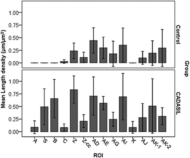
Figure 4.

Distribution and density of SMI32-positive axons. In general, cerebral autosomal dominant arteriopathy with subcortical infarcts and leukoencephalopathy (CADASIL) had more axonal staining than controls especially in frontal lobes. The density of abnormal axons correlated with disease duration (see Table 4). Z-cc: corpus callosum area in block Z; ROI: region of interest.
