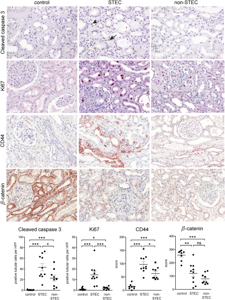
Figure 5.

Immunohistochemical findings of the STEC-associated acute tubular damage. Expression of cleaved caspase 3, Ki67, β-catenin and CD44 were studied by immunohistochemistry in STEC-infected patients (n = 10) and compared to normal kidney tissue from tumour nephrectomies (control, n = 8) and to kidney biopsies with TMA due to a STEC-unrelated aetiology (non-STEC, n = 9). The acute tubular damage was associated with a significant increase in apoptosis (visualized by cleaved caspase 3), reactive proliferation (visualized by Ki67) and elevated expression of CD44 in both STEC and non-STEC groups. However, for all three parameters, the increase was significantly higher in the STEC than in the non-STEC group. In the STEC group, apoptosis (arrow) and mitosis (arrowhead) could be seen in tubular epithelial cells; β-catenin was significantly down-regulated in renal tubular epithelium of both STEC and non-STEC patients, to a similar extent; magnification = ×100. In the case of immunohistochemistry for cleaved caspase 3 and Ki67, the number of positive tubular cells was counted and expressed as average cell number/high power field (HPF; ×40 objective, ×10 ocular). For evaluation of the immunohistochemistry for β-catenin and CD44, semiquantitative scoring of the staining intensity of tubular cells in each HPF of the biopsy was performed; in the non-STEC group, individuals > 45 years are depicted as squares. Statistical differences were tested by two-tailed Mann–Whitney test; ns, non-significant; *p < 0.05; **p < 0.01; ***p < 0.001
