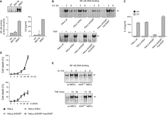
HeLa cells were left untreated (ctrl) or were infected with non-invasive Shigella strain BS176 or the invasive strain M90T (MOI 30). NF-κB (p65) DNA binding activity was analyzed by EMSA (upper panel) or ELISA (lower panel) 2 h post-infection (p.i.). IL-8 secretion was measured by ELISA in supernatants of cells 6 h p.i. (lower right panel). Data are presented as mean ± SEM (n = 3).
HeLa wt, HeLa shScr, HeLa shXIAP and HeLa shXIAP-mycXIAP were left untreated (–) or were infected with Shigella M90T (MOI 30) (upper panel) or stimulated with TNF (10 ng/ml, 30 min) (lower panel). NF-κB (p65) DNA binding activity was analyzed by EMSA at the indicated time points p.i..
HeLa wt, HeLa shScr, HeLa shXIAP and HeLa shXIAP-mycXIAP were left untreated (ctrl) or were infected with Shigella M90T (MOI 30). IL-8 secretion was monitored by ELISA in supernatants of cells 6 h p.i.. A representative experiment of three independent experiments is shown. Data are presented as mean ± SEM (n = 3).
HeLa wt, HeLa shScr, HeLa shXIAP and HeLa shXIAP-mycXIAP were infected with Shigella M90T with MOI 30 (left panel) or the indicated MOI (right panel). Cell death was determined at the indicated time points p.i. by trypan blue exclusion. Data are presented as mean ± SD (n = 3).
MEFs isolated from wt or XIAP−/− mice were infected with Shigella M90T (MOI 30) (upper panel) or were treated with TNF (10 ng/ml, 30 min) (lower panel). NF-κB (p65) DNA binding activity was analyzed by EMSA at the indicated time points p.i..

